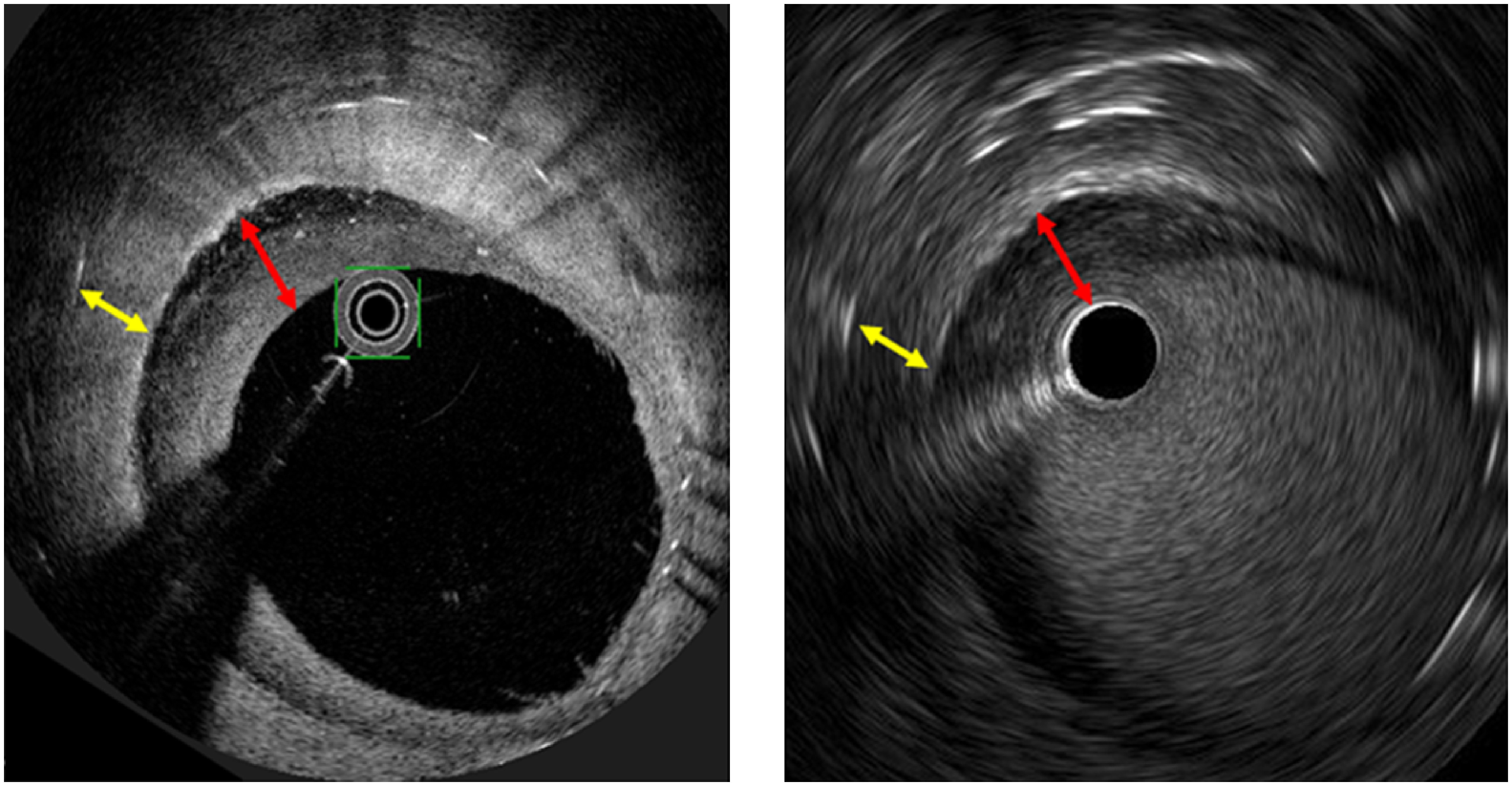
Fig. 4 Comparison of the multi-layered ISR pattern between OFDI and IVUS. (A) OFDI image, a superficial layer (red arrow), and a deep layer (yellow arrow). (B) IVUS image, a superficial hypoechoic layer (red arrow), and a deep layer (yellow arrow).
ISR: in-stent restenosis; OFDI: optical frequency domain imaging; IVUS: intravascular ultrasound

