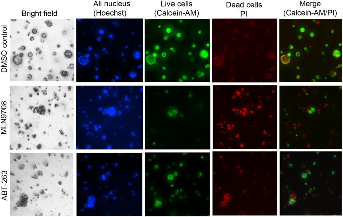
Figure 5.
Imaging analysis of cell fate of organoids in a 384-well plate format. Organoids growing in a 384-well plate (4 days) were treated with DMSO, MLN9708, or ABT-263 (5 µM) for 3 days. A combination of dyes diluted in PBS containing Hoechst 33342, Calcein-AM, and PI was added and incubated for 30 min. The bright field and fluorescence images were captured with the ImageXpressmicro automated imaging system using Z-stack. The representative merged images from Z-stack for DMSO control, MLN9708, and ABT-263 treatment are shown.

