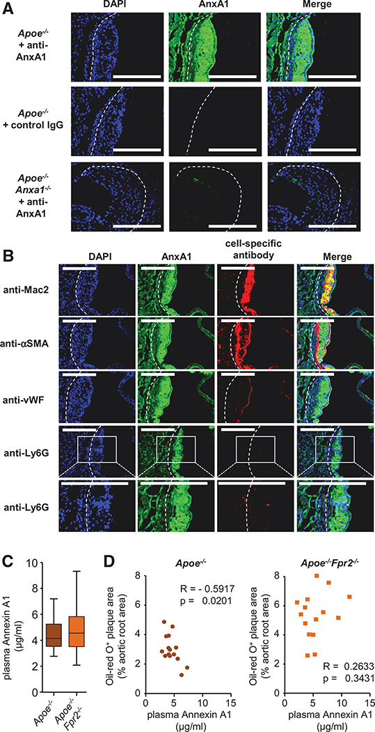
Figure 2. Plasma annexin A1 (AnxA1) negatively correlates with atherosclerotic lesion sizes.
A, Apoe−/− and Apoe−/−Anxa1−/− mice were fed a high-fat diet for 4 weeks, and aortic root sections were stained with an antibody to AnxA1 or an isotype control antibody. B, Apoe−/− mice were fed a high-fat diet (HFD) for 4 weeks and AnxA1 was costained with markers for macrophages (anti-Mac2), smooth muscle cells (anti-αSMA), endothelial cells (anti-von Willebrand Factor [vWF]), or neutrophils (anti-Ly6G) in aortic root sections. Scale bar, 100 μm. C and D, Apoe−/− and Apoe−/−Fpr2−/− mice were fed a HFD for 4 weeks and AnxA1 was quantified in the plasma (B). Correlation between aortic root lesion sizes and plasma AnxA1 levels in Apoe−/− and Apoe−/−Fpr2−/− mice (Pearson correlation; D). DAPI indicates 4’,6-diamidino-2-phenylindole.

