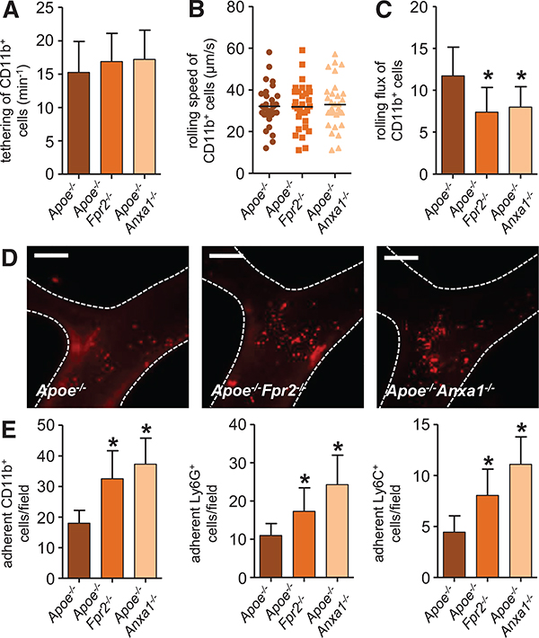
Figure 3. Annexin A1-formyl peptide receptor 2 axis prevents arterial myeloid cell recruitment.
Apoe−/−, Apoe−/−Fpr2−/−, and Apoe−/−Anxa1−/− mice were fed a high-fat diet for 4 weeks, and intravital microscopy of the carotid artery was performed to assess luminal leukocyte endothelial interactions. Myeloid cell subsets were identified by intravenous injection of antibodies to Ly6G, Ly6C, and CD11b 10 minutes before recording. Displayed are quantification of tethering (A), rolling speed (B), and rolling flux (C) of CD11b+ cells. Number of adherent cells (E) is displayed for CD11b+ cells (left), Ly6G+ cells (middle), and Ly6C+ cells (right). Adhesion of CD11b+ myeloid cells to carotid arteries of Apoe−/−, Apoe−/−Fpr2−/−, and Apoe−/−Anxa1−/− mice is depicted in D. Scale bar, 100 μm. *P<0.05 compared with Apoe−/− mice. Experiments were performed 3× independently with a total of ≥15 mice. Data were analyzed using 1-way ANOVA with Dunnett post test.

