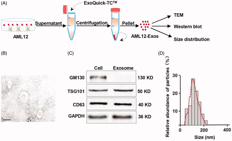
Figure 1.
Isolation and identification of AML12 cell-derived exosomes. (A) Schematic representation of the exosomes isolation procedure; (B) representative TEM (transmission electron microscope) image of the isolated exosomes (scale bar = 100 nm); (C) western blot analysis of the expression of exosomal markers GM130, TSG101 and CD63 in the isolated exosomes and the parental cells. GAPDH served as the loading control; (D) size distribution of the isolated exosomes.

