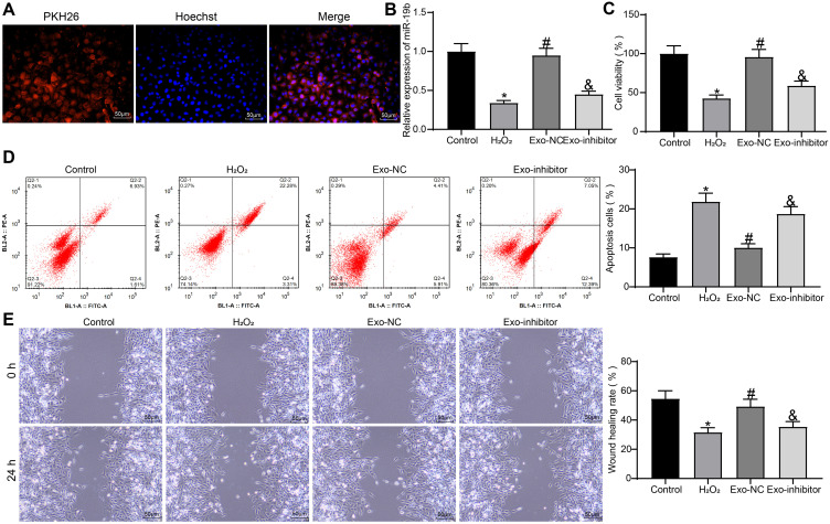
Figure 6.
Exosomal miR-19b promotes HSF growth and migration. (A) HSF uptake of exosomes. (B) Expression of miR-19b in HSF cells was detected by RT-qPCR (one-way ANOVA, *P < 0.05 vs the Control group; #P < 0.05 vs the H2O2 group; &P < 0.05 vs the Exo-NC group). (C) MTT assay to detect the viability of cells (one-way ANOVA, *P < 0.05 vs the Control group; #P < 0.05 vs the H2O2 group; &P < 0.05 vs the Exo-NC group). (D) Flow cytometry to detect apoptosis rate of cells (one-way ANOVA, *P < 0.05 vs the Control group; #P < 0.05 vs the H2O2 group; &P < 0.05 vs the Exo-NC group). (E) Wound healing detected the migration ability of HSF cells in each group (one-way ANOVA, *P < 0.05 vs the Control group; #P < 0.05 vs the H2O2 group; &P < 0.05 vs the Exo-NC group).

