
Figure 5.
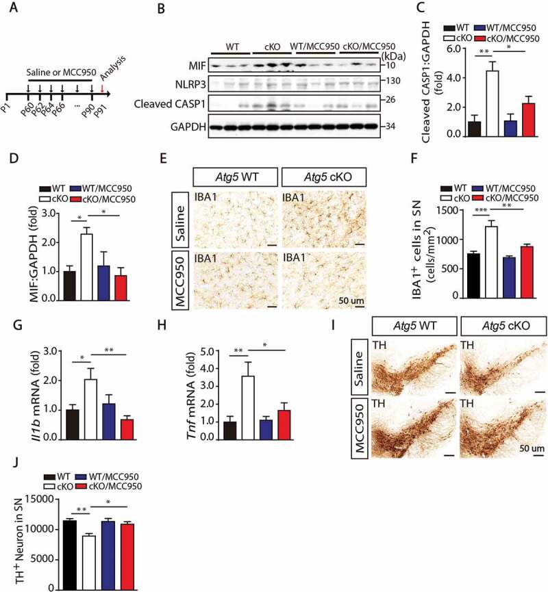
Inhibition of NLRP3 activation rescued Atg5 defect induced microglial activation and neurons loss. (A) Model of administration of MCC950 to block NLRP3 activation and check the microglial activation and neurons loss. Two-month-old Atg5 WT and cKO mice received intraperitoneal injections of sterile PBS (control) or MCC950 (10 mg/kg in PBS) every second day for 1 month. (B) Immunoblotting analysis of MIF, NLRP3, cleaved CASP1 and GAPDH in the SN from Atg5 WT group, Atg5 cKO group, Atg5 WT with MCC950 group and Atg5 cKO with MCC950 group mice. (C and D) Quantitative data of cleaved CASP1 and MIF levels in the SN from these 4 groups of mice as indicated. (E) Immunohistochemical staining for IBA1 in the SN from these 4 groups of mice. (F) Quantitative data of IBA1 positive cells in the SN from these 4 groups of mice as indicated (N = 6 or N = 8). (G and H) Expression of Il1b and Tnf in the SN from these 4 groups of mice as indicated. (I) Immunohistochemical staining for TH in the SN in these 4 groups of mice as indicated. (J) Quantitative data of TH-positive neurons in the SN in these 4 groups of mice as indicated (N = 4 for each group)