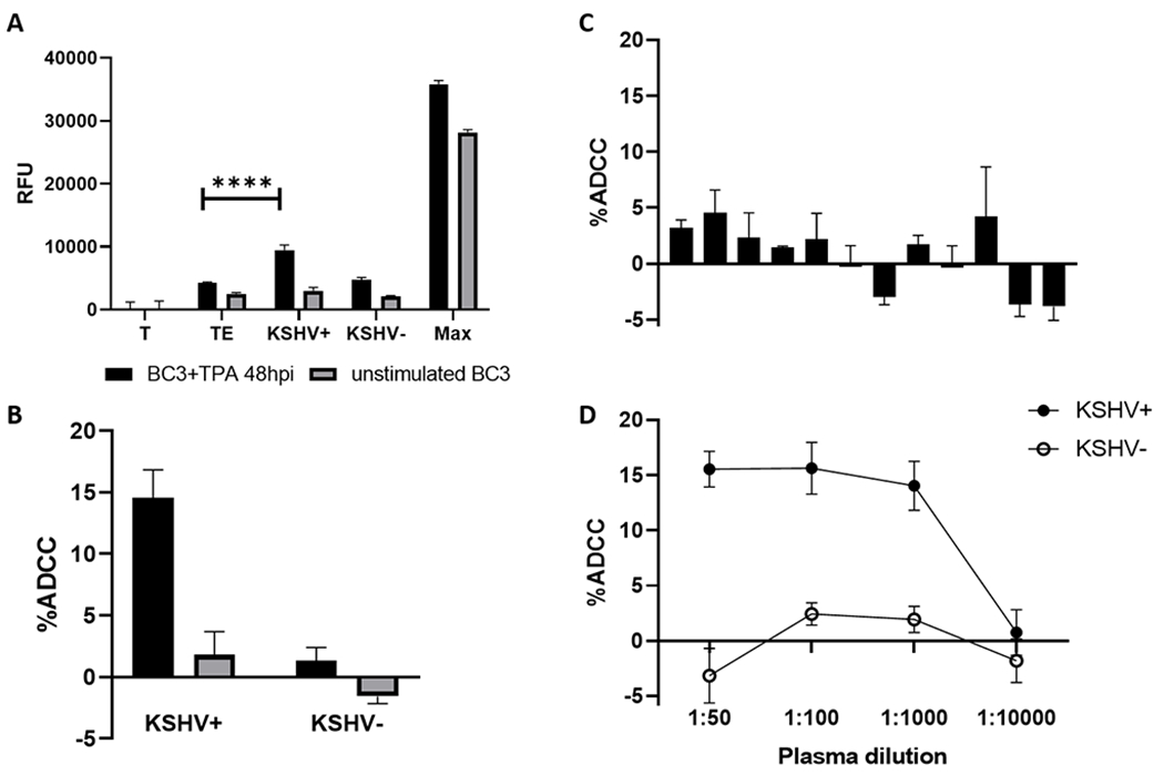
Figure 1.

Validation of a KSHV specific ADCC assay. (A) Relative fluorescent units (RFU) of supernatant from wells containing targets only (T), target and effectors (TE), targets and effectors plus KSHV seropositive plasma (KSHV+), targets and effectors plus KSHV seronegative plasma (KSHV−), and target cells plus detergent (Max) for BC3 cell stimulated with PMA (black bars) or unstimulated (gray bars). (B) Percent ADCC was calculated as (Experimental-TE)/(Max-T)*100. (C) ADCC activity for 12 KSHV seronegative individuals. (D) ADCC activity at varying dilutions of KSHV seropositive and seronegative plasma. All data shown is the mean and standard deviation from the centroid three of five experimental replicates. Significant differences denoted: **** p<0.0001
