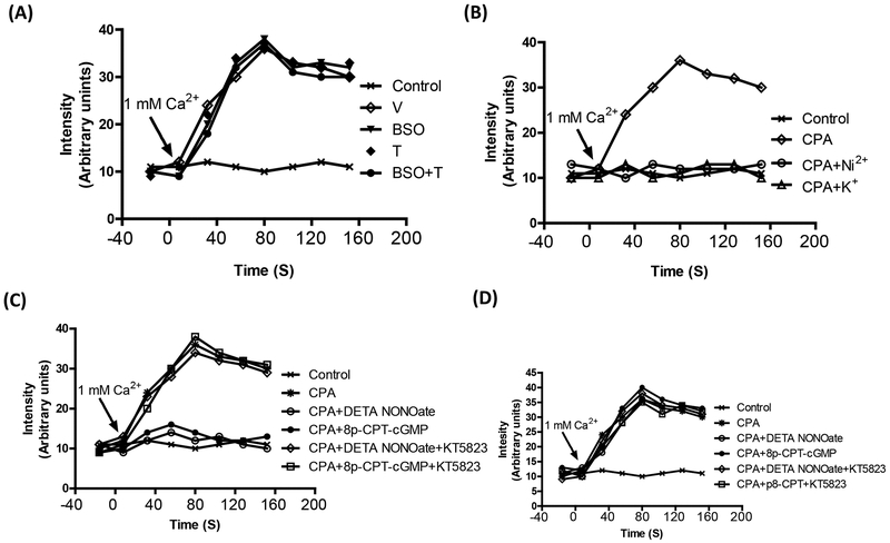
Figure 6. Cyclopiazonic acid (CPA)-induced increase in intracellular Ca2+.
A, Mesenteric vessels, from vehicle (V), L-buthionine sulfoximine (BSO), tempol (T) and BSO + T -treated rats, was incubated with 10 μM CPA in Ca2+ free buffer followed by change to 1mM Ca2+ buffer. Ca2+ influx was monitored by Fluo2/AM fluorescence. B, Ca2+ influx in mesenteric vessel from vehicle-treated rats incubated with CPA in the presence and absence of Ni2+ (3 mM) which competes with Ca and blocks its entry and 80 mM K+ for membrane depolarization. C, Ca2+ influx in mesenteric vessels from vehicle-treated rats incubated with CPA in the presence and absence diethylenetriamine NONOate (DETA NONOate), 8-para-chlorophenylthio-cGMP (8p-CPT-cGMP) or KT5823 (1 μM, a PKG inhibitor). D, Ca2+ influx in mesenteric vessel from BSO-treated rats incubated with CPA in the presence and absence DETA NONOate, 8p-CPT-cGMP or KT5823. A single experiment is shown, representative of 6–8 individual experiments performed in triplicate.

