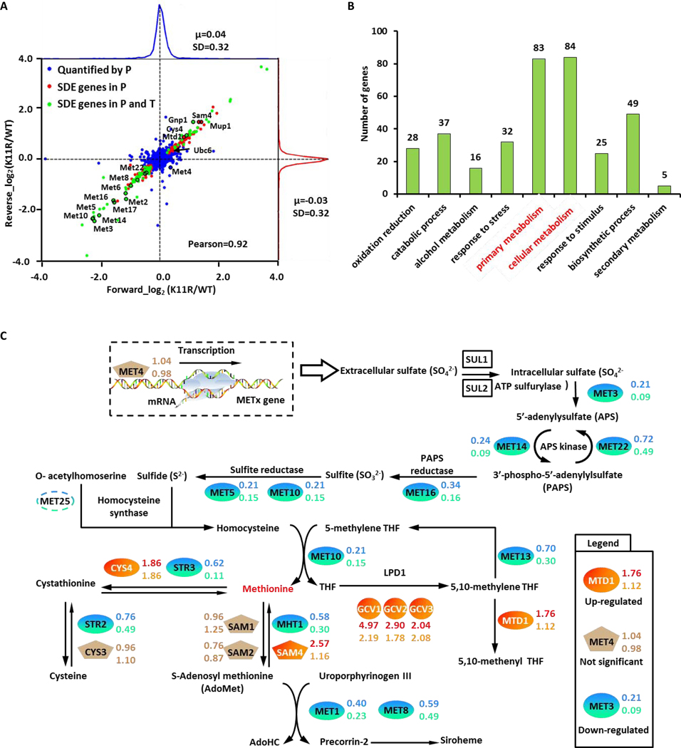
Figure 2. Potential protein substrates of K11-linked ubiquitin chains.
(A) Log2 (K11R/WT) ratio distributions of quantified proteins. Blue, unchanged proteins; red, significantly changed proteins identified through proteomics; green, significantly changed proteins identified through proteomics and transcriptomics. SDE gene stood for the significant different expressed gene.
(B) Gene ontology analysis of potentially K11-linked ubiquitylated substrates, namely the upregulated protein in K11R datasets.
(C) Members of the methionine biosynthesis pathway are colored according to fold change. The protein and mRNA ratios of K11R to WT were determined for each protein-coding gene.

