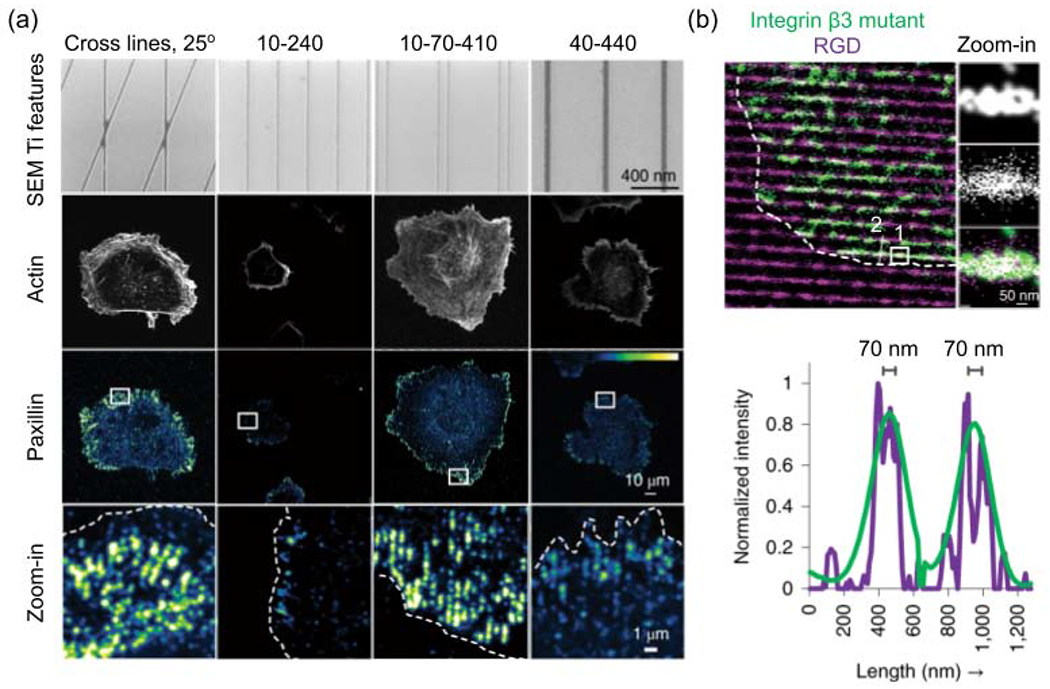
Figure 2.

Cell adhesion on nanoline arrays. (a) Representative images of cell adhesion and spreading on different nanoline patterns, which are systematically named by the geometric parameters of features. For line pairs, the first number is line width, the second is internal edge-to-edge spacing between the two lines in a pair, and the third is edge-to-edge spacing between adjacent line pairs, in a unit of nm. Fluorescent protein (mApple)-tagged paxillin indicates the cell adhesion. (b) Integrin beta3 binds to nanolines coated with the RGD ligand. The figure is adapted with permission from [42], Springer Nature.
