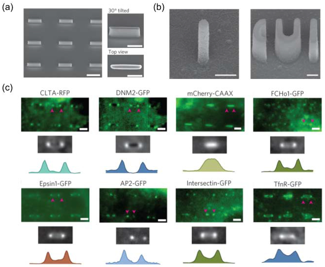
Figure 4.

Nanotopography enhances clathrin-mediated endocytosis in a curvature-dependent manner. (a) SEM images of a quartz nanobar array showing individual nanobars of 150 nm width, 2 μm length, 1 μm height and 5 μm pitch. Scale bar, 2 μm. Nanobar structure locally induces high curvature at the two ends and zero curvature in the middle. (b) SEM images of quartz nanopillar and nanoCUI structures. Scale bars, 500 nm. [73] (c) High-magnification fluorescence images, scale bars, 2 μm. They show the bar-end/high-curvature distributions of clathrin(CLTA), dynamin2 (DYM2), mCherry-CAAX, FCHo1, Epsin1, AP2, Intersection and TfnR on six nanobars. Other endocytic proteins involved in different stages of endocytosis were also shown in the work. (a,c) are adapted with permission from [72], Springer Nature. (b) is adapted with permission from [73], Springer Nature.
