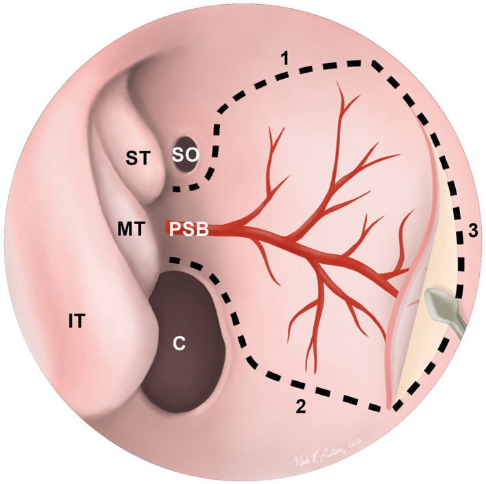
FIGURE 4.

Endoscopic view of a nasoseptal flap (NSF) in the right nasal cavity. Dotted lines depict the superior (1), inferior (2), and anterior (3) incisions of the NSF. The inferior incision can be extended laterally onto the nasal floor to maximize the surface area of the graft. Once elevated, the flap can be tucked into the nasopharynx or maxillary sinus until it is needed for reconstruction. C, choana; IT, inferior turbinate; MT, middle turbinate; PSB, posterior septal branch of the sphenopalatine artery; SO, sphenoid ostium; ST, superior turbinate
