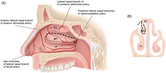
FIGURE 5.

Sagittal, A and coronal, B, views of the middle turbinate flap (MTF) surgical anatomy. The MTF is pedicled on the posterior lateral nasal branches of the sphenopalatine artery. Borders of the flap are depicted by with dotted lines, A. The flap is raised from both the medial and lateral aspects of the turbinate, B. IT, inferior turbinate; MT, middle turbinate; ST: superior turbinate
