FIGURE 3.

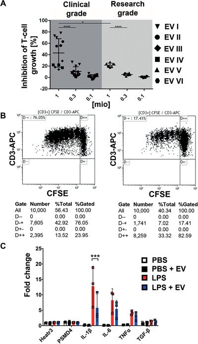
Immunomodulatory potential of mesenchymal stromal cell‐derived extracellular vesicles (MSC‐EVs). A, Four clinical‐grade (I‐IV) and two research‐grade (V and VI) batches of MSC‐EVs were tested for their capacity to inhibit phytohemagglutinin (PHA)‐induced T‐cell proliferation in dilution series as indicated (1, 0.3, 0.1 million particles per 5 × 10⁵ mononuclear cells). To determine the percentage of inhibition of carboxyfluorescein succinimidyl ester (CFSE)‐labeled CD3 T‐cell proliferation, samples were analyzed by flow cytometry in triplicates, and one‐way ANOVA was used for statistical analysis (*P < .05; ****P < .0001). Results are shown as mean ± standard deviation. B, Representative dot plots show CD3+ T‐cell proliferation kinetics without inhibition in the absence (left), or with inhibition in the presence of MSC‐EVs (right), respectively. For normalization, the standard stimulation (PHA only, left dot plot, upper left quadrant) was assigned to a value of 100% and the percentage of inhibition was calculated with 10 000 CD3+ T cells gated per analysis. C, The treatment of BV‐2 microglial cells with lipopolysaccharides (LPS) rapidly upregulates the gene expression of the proinflammatory cytokines IL‐1β, IL‐6, and TNFα. The presence of MSC‐EVs significantly reduced the induction of IL‐1β expression in BV‐2 cells in response to LPS (fold change is normalized to the endogenous controls Heatr3 and PSMD4)
