FIGURE 5.

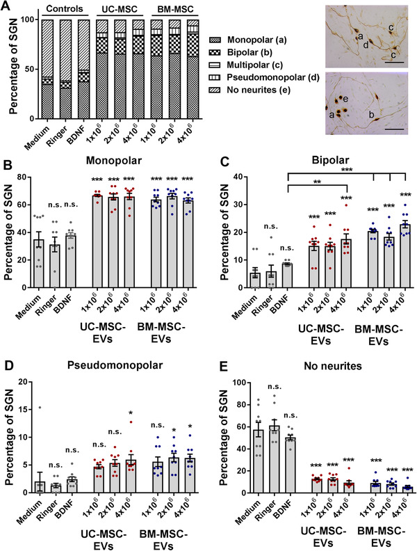
Mesenchymal stromal cell‐derived extracellular vesicles (MSC‐EVs) alter the morphology of spiral ganglion neurons (SGN). A, The various morphologies of SGN treated with research‐grade EV preparations derived from umbilical cord and bone marrow‐derived mesenchymal stromal cells (UC‐ and BM‐MSC) are depicted in an overview graph (left). Representative images of SGN with mono‐, bi‐, multi‐, pseudomonopolar morphology, and SGN lacking neurites are shown (right) and designated as (a) to (e). B, The percentage of monopolar neurons increases in SGN treated with all EV preparations and concentrations. C, The highest concentration of the UC‐MSC‐EVs and all tested BM‐MSC‐EV doses clearly increase the percentage of bipolar neurons in comparison to brain‐derived neurotrophic factor (BDNF). D, The percentage of pseudomonopolar neurons increases after the treatment with high concentrations of EVs. E, The number of SGN lacking neurites is significantly reduced in the presence of MSC‐EVs. Scale bar: 100 μm; number of experiments: N = 3, number of replicates per experiment: n = 3. ***P < .001; **P < .01; *P < .05; significance levels indicated above individual bars show comparison with medium (negative control), significance levels in comparison to the positive control BDNF are separately depicted by horizontal lines. Each data point represents the determined percentage of SGN in a single well (B‐E)
