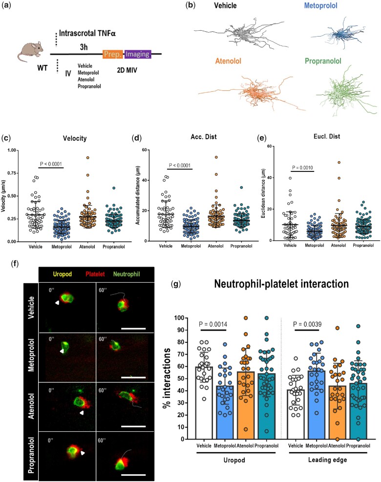
Figure 4.
Metoprolol has a particular disruptive effect on neutrophil dynamics in vivo. (A) Experimental scheme for 2D intravital microscopy (IVM) of neutrophil motility in inflamed cremaster muscle. (B) Representative tracks of crawling neutrophils within inflamed vessels of mice treated with vehicle, metoprolol, atenolol, or propranolol. (C–E) Two-dimensional intravascular motility parameters: velocity (µm/s), accumulated distance (µm), and euclidean distance (µm); n = 52–89 cells from 5 to 6 mice per condition. (F) Representative time-lapse images of platelets (CD41+ cells, red) with the polarized neutrophil uropod (CD62L+ domain, yellow) or leading edge (Ly6G+ domain, green) in the different conditions. Arrowheads indicate interactions with the uropod domain, and dotted lines indicate displacement of the neutrophil over 60s. (G) Percentage of platelet interactions with the neutrophil uropod or leading edge; n = 24–37 cells from three mice per condition. Data are presented as means ± SD.

