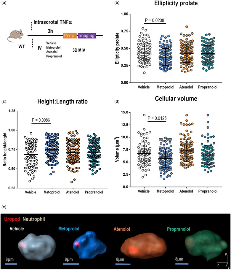
Figure 5.
Metoprolol alters neutrophil polarized morphology. (A) Experimental scheme for 3D intravital microscopy (IVM) of neutrophil morphology in inflamed cremaster muscle. (B–D) Three-dimensional intravascular cell morphology parameters: ellipticity prolate, height: length ratio, and volume. n = 75–118 cells from 3 to 4 mice per condition. Data are presented as mean ± SD. (E) Representative 3D reconstructions of polarized neutrophils (uropod, red) within live cremaster vessels of mice treated with vehicle (grey), metoprolol (blue), atenolol (orange), or propranolol (green).

