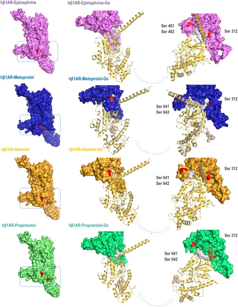
Figure 7.
Ligand-human β1AR-Gs protein α subunit conformational changes. Modelling of the binding of the Gsα subunit to the complexes established upon docking of the different ligands (epinephrine, purple; metoprolol, blue; atenolol, orange; and propranolol, green) to the human β1AR.The binding of the Gsα subunit to the metoprolol–β1AR complex exposes phosphorylation sites in the receptor potentially triggering GRK/β-arrestin signalling cascade (see text). Red areas indicate Ser phosphorylation sites.

