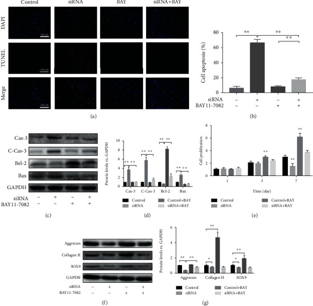
Figure 6.

LMP-1 silencing increased apoptosis of NPCs by activating the NF-κB signaling pathway. (a) TUNEL method was performed to measure apoptosis of NPCs (red), and the results were observed by fluorescence. Nuclei (blue) were stained by DAPI. (b) Cell apoptosis was quantified according to the results of TUNEL. ∗∗p < 0.01. (c) The protein expression levels of caspase-3, cleaved-caspased-3, Bcl-2, and Bax of NPCs were measured by western blotting analysis on day 3. (d) The protein expression levels of caspase-3, cleaved-caspased-3, Bcl-2, and Bax of NPCs were quantified. ∗∗p < 0.01. (e) Cell proliferation on each group was measured by CCK-8 assay on days 1, 3, and 7. ∗∗p < 0.01 vs. the control group. (f) The protein expression levels of aggrecan, SOX9, and collagen II of NPCs in each group were measured on day 14 by western blotting analysis. (g) The protein expression levels of aggrecan, SOX9, and collagen II of NPCs were quantified. ∗p < 0.05, ∗∗p < 0.01. BAY11-7082 was used as a specific inhibitor of the NF-κB signaling pathway. Data represent mean ± SD; scale bar = 200 μm.
