
Figure 1.
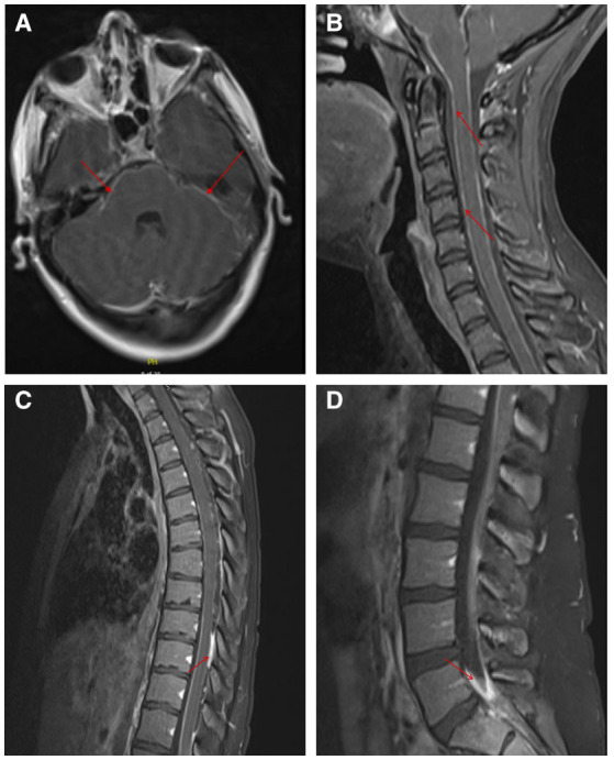
Leptomeningeal enhancement demonstrated on MRI with and without contrast of the brain and spinal cord. MRI brain axial image showing leptomeningeal enhancement of the posterior fossa and the visualized proximal spinal cord (A). Leptomeningeal enhancement was noted in sagittal images of the cervical spine (B), thoracic spine (C), and lumbar spine (D). Areas of hyperintensity are denoted by the red arrows.