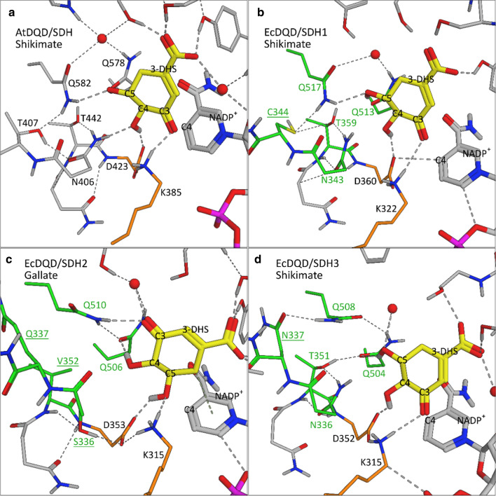
Fig. 9.
Model structures of the SDH ternary complex of AtDQD/SDH (a) and EcDQD/SDH1–3 (b, c, d). Key substrate-binding residues are in gray (AtDQD/SDH) or green (EcDQD/SDHs). Substituted amino acids are underlined. The catalytic dyad is marked in orange. The substrate 3-DHS (yellow, bold) is presented in the orientation required for shikimate (a, b, d) or gallate (c) formation. Water molecules are represented by red spheres. Hydrogen bridges are indicated as dashed lines

