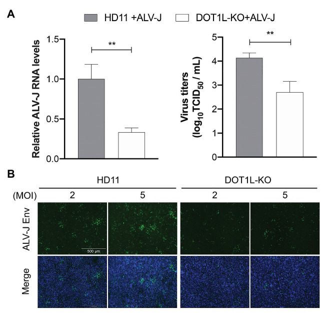
Figure 4.

Knockout of DOT1L inhibits ALV-J replication. (A) HD11 and DOT1L-KO cells were infected with ALV-J (MOI 2). After 48 h, the viral RNA levels were measured by qRT-PCR. GAPDH mRNA level was measured as an internal control (left panel). The supernatants were collected to measure the viral titers by a standard TCID50 method (right panel). (B) Immunofluorescence analyses of ALV-J Env protein. HD11 cells and DOT1L-KO cells were infected with ALV-J at indicated MOI for 48 h, the cells were then visualized with the inverted fluorescence microscope and a specific antibody to the ALV-J Env protein (green), and nuclei were stained with 4', 6-diamidino-2-phenylindole (DAPI; blue). Images show a representative image. All the data were shown as mean ± SD (error bars) from three independent experiments. **p < 0.01; unpaired, two-tailed Student’s t-test.
