FIGURE 2.
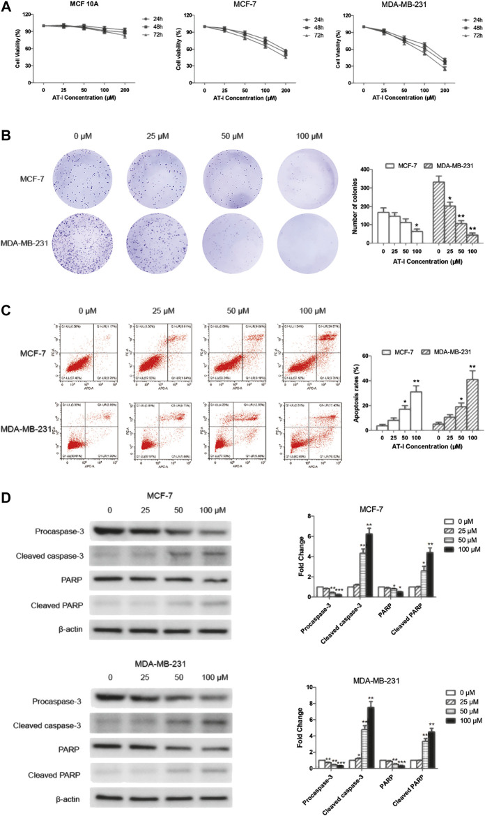
AT-I inhibited cell growth and induced apoptosis in breast cancer cells. (A) MCF 10A, MCF-7 and MDA-MB-231 cells were treated with AT-I (0–200 µM) for 24, 48 and 72 h to determine cell viability. (B) MCF-7 and MDA-MB-231 cells were treated with AT-I (0–100 µM) for 48 h and then applied for colony formation. The number of colonies was counted after 14 days. (C) MCF-7 and MDA-MB-231 cells apoptosis assay after treatment with AT-I (0–100 µM) for 48 h. (D) The effects of AT-I on the expression of procaspase-3, cleaved caspase-3, PARP and cleaved PARP were measured by western blot assay. Significant differences between different groups were indicated as *p < 0.05, **p < 0.01, ***p < 0.001, vs. the 0 μM control group, n = 3.

