FIGURE 1.

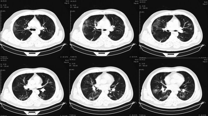
Chest CT images of the patient. Ground‐glass opacities are seen in both lungs, predominantly in lower zones. Multiple atelectasis is also seen in both lower zones

Chest CT images of the patient. Ground‐glass opacities are seen in both lungs, predominantly in lower zones. Multiple atelectasis is also seen in both lower zones