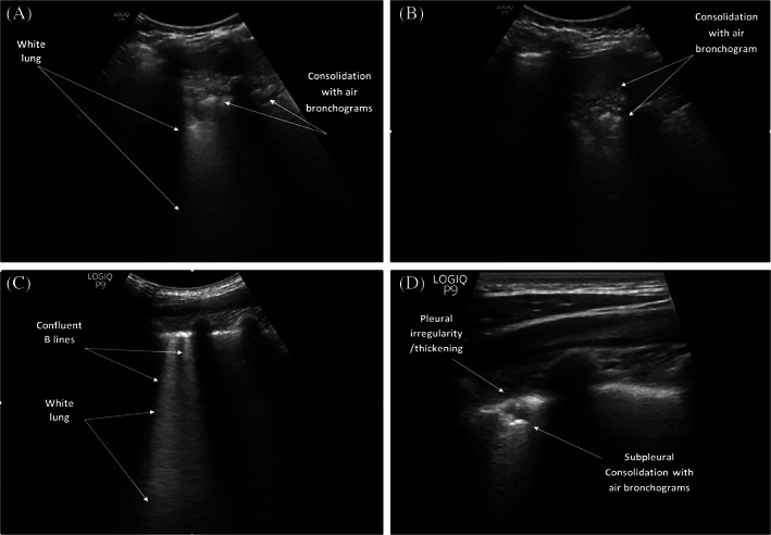
FIGURE 3.

A, Lung ultrasound (LUS) shows white lung appearance and consolidations with air bronchograms. B, LUS shows consolidations with air bronchograms. C, Confluent B lines and white lung appearance. D, LUS image of pleural thickening/irregularity and consolidations with air bronchograms obtained using a linear transducer
