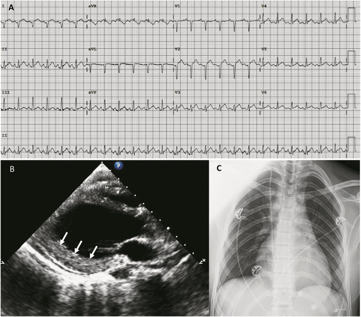
Figure 1.

Electrocardiogram (EKG), transthoracic echocardiogram (TTE), and chest X‐ray at admission. (A) EKG showing sinus tachycardia, inframillimetric ST segment elevation, and PR depression. (B) Parasternal long axis view of left ventricular (LV) demonstrating non‐dilated LV (end‐diastolic diameter 42 mm) and wall thickening of inferolateral wall (13 mm) with granulated myocardial appearance (white arrows). (C) Absence of lung infiltrates.
