Figure 3.

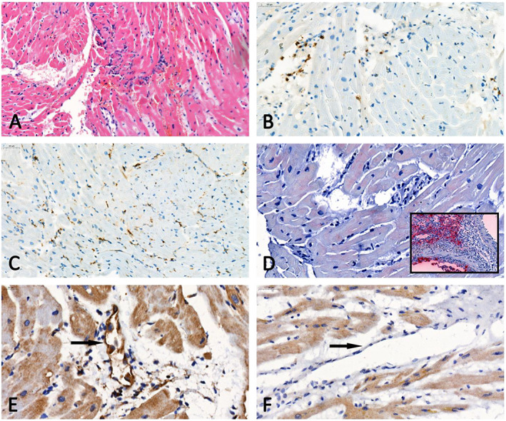
Optical microscopy of heart tissue. (A) Haematoxylin–eosin section showing a low density of mononuclear inflammatory cells (open arrows) in the absence of myocyte degeneration or necrosis. (B, C) Representative examples of CD3 (B) and CD68 (C) immunostaining demonstrating < 14 T lymphocytes or macrophages/mm2. (D) Absence of immunoreactivity for the viral nucleocapsid protein in the biopsy of our patient, which contrasts with the intense staining in the positive control [inset; lung tissue specimen of a severe acute respiratory syndrome coronavirus 2 (SARS‐CoV‐2) infected hamster]. (E, F) Immunohistology sections for fractalkine expression showing respectively an endothelial immunoreactivity in the biopsy of our patient (E) and no staining in the myocardium of a patient who died from a coronavirus disease 2019 (COVID‐19)‐unrelated cause (F).
