
Figure 2.
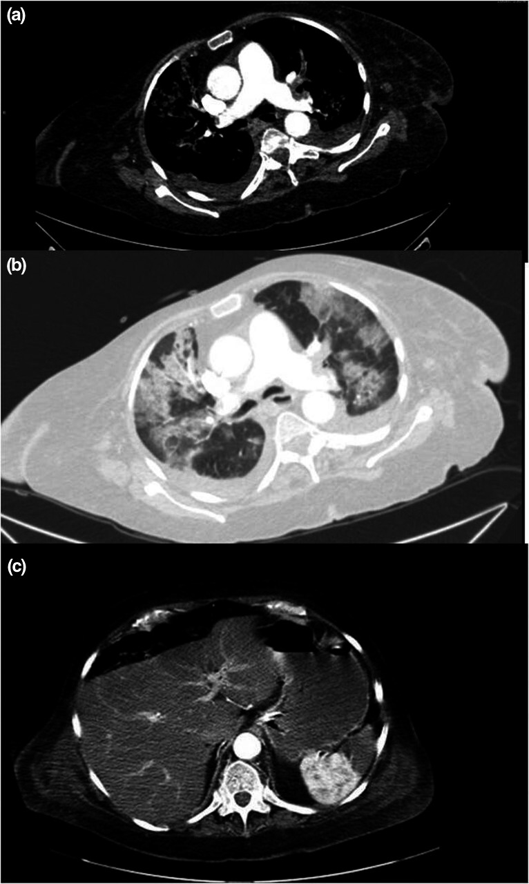
(a) Computed tomography angiography showed massive bilateral pulmonary thromboembolism. (b) Computed tomography showed bilateral and peripheral ground‐glass opacities with consolidations. (c) Computed tomography angiography showed a hypodense lesion with triangular morphology in the spleen, compatible with splenic infarction