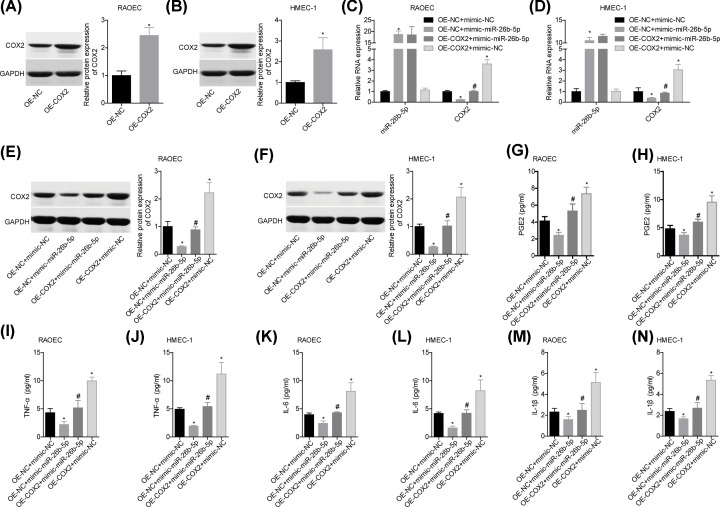
Figure 4. miR-26b-5p repressed inflammatory responses via targeting COX2 in vitro.
(A,B) Following 48 h of cell transfection with OE-NC/OE-COX2, RAOEC and HMEC-1 cells were collected for Western blotting assay to detect the protein level of COX2. Then, cells in OE-NC+mimic-NC, OE-NC+mimic-miR-26b-5p, OE-COX2+mimic-miR-26b-5p and OE-COX2+mimic-NC groups were submitted to the following assays. (C,D) The levels of miR-26b-5p and COX2 mRNA in cells were tested by using qPCR. (E,F) The protein level of COX2 was determined by Western blotting. (G–N) The levels of PGE2, TNF-α, IL-6 and IL-1β in the supernatants of cells were measured by ELISA (n=3, *P<0.05, vs. OE-NC+mimic-NC group; #P<0.05, vs. OE-NC+mimic-miR-26b-5p group).

