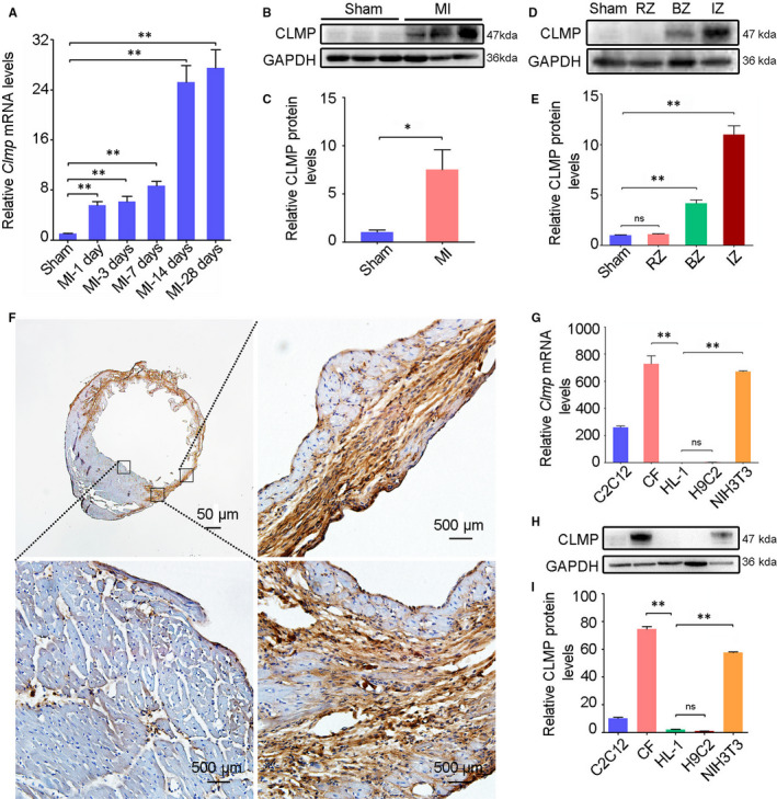
FIGURE 1.

Clmp is highly expressed in cardiac fibroblasts and induced in the ischaemic heart. A, The mRNA expression of Clmp was significantly increased in cardiac tissue of the wild‐type mice on the indicated days post‐MI (n = 3). B, The representative Western blot imaging of CLMP protein in the sham‐operated and infarcted heart tissues (week 2 after MI). C, The relative densitometric quantification of panel B showed higher expression level of CLMP protein in the ischaemic heart (n = 3). D, The representative Western blot imaging of CLMP protein in the heart lysates from the sham, as well as the non‐ischaemia zone, border zone and infarct zone of the MI heart. E, The relative densitometric quantification of panel D showed the highest expression level of CLMP protein in the IZ of the MI heart(n = 3). F, The representative immunohistochemistry imaging showed significantly increased expression of CLMP in the MI heart. Scale bar indicates 50 μm. G, qRT‐PCR analysis showed the Clmp expression in different fibroblasts (CFs: cardiac fibroblasts; NIH3T3) and myogenic cells (HL‐1, C2C12, H9C2). H, Western blot showed abundant expression of CLMP protein in the fibroblasts rather than myogenic cells. I, The relative densitometric quantification of CLMP protein in panel H. All data are presented as the mean ± SEM. Student's t test or one‐way ANOVA; *P < .05; **P < .01; ns, not significant
