
FIGURE 3.
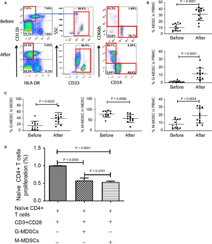
Glucocorticoid treatment amplified the percentages of MDSC and the two subsets in PBMC of relapsing MS patients. A, Staining profiles of MDSC (CD11b+CD33+HLA‐DR−), M‐MDSC (CD11b+CD33+HLA‐DR−CD14+CD66b−) and G‐MDSC (CD11b+CD33+HLA‐DR−CD14−CD66b+) from a representative MS patient before and after methylprednisolone impulse treatment. B, Percentages of MDSC (P < 0.0001), G‐MDSC (P = 0.0001) and M‐MDSC (P = 0.0024) in PBMC of MS patients (n = 12) before and after treatment. C, Percentages of G‐MDSC (P = 0.0035) and M‐MDSC (P = 0.0096) in MDSC of PBMC from MS patients (n = 12) before and after treatment. D, Naïve human CD4+ T cells were stimulated by anti‐CD3/CD28 in the absence or presence of MDSC from MS patients for 3 d (G‐MDSC, P = 0.0005, n = 3; M‐MDSC, P < 0.0001, n = 3; G‐MDSC vs M‐MDSC, P = 0.3701, n = 3), and T‐cell proliferation was determined by measuring BrdU incorporation (Brdu was added 6 h prior to cell harvest)