Figure 3.

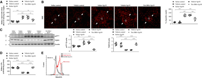
Tris DBA reduced oxidative stress and enhanced ROS activity in IgAN mice. A, Superoxide anion in renal tissues; B, DHE staining on renal tissues and the quantified results of DHE staining. Original magnification, 400X. Arrow indicates glomerular area; C, the protein level of NADPHp47 and NQO1 in Western blot analysis in renal tissue and semiquantitative analysis; D, renal GPx level in ELISA. Bars show the mean ± SEM results in 7 mice per group. E, Mitochondrial ROS levels in BMDMs measured by MitoSOX after Tris DBA treatment, priming with IgA‐ICs for 5.5 h and stimulation with ATP. The data are expressed as the means ± SEM for three separate experiments. DHE—Dihydroethidium; NADPHp47—NADPH oxidase subunit p47 (phox) and NQO1—NADPH:quinone oxidoreductase 1; GPx—Glutathione peroxidase. **P < .01, ***P < .005, ****P < .001
