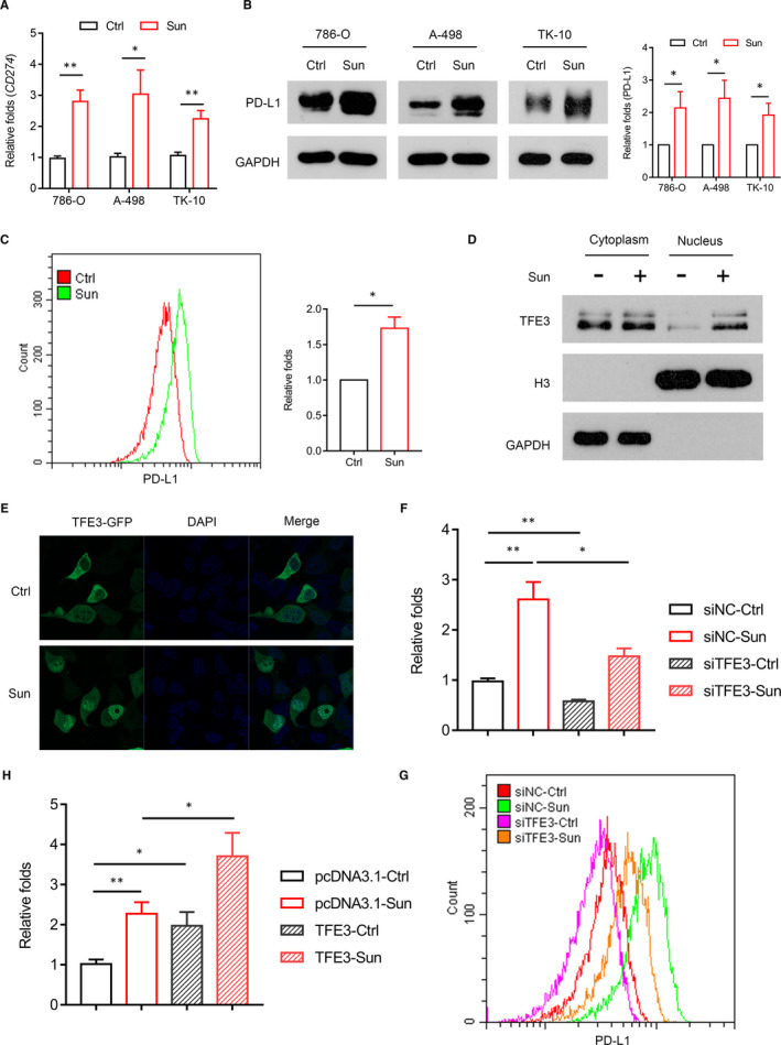
Figure 4.

Sunitinib enhances PD‐L1 expression via activation of TFE3 in ccRCC cells. A, PD‐L1 expression was analysed by qPCR in multiple cells (786‐O, A498, TK‐10) with sunitinib treatment. B, PD‐L1 expression was analysed by Western blots in multiple cells (786‐O, A498, TK‐10) with sunitinib treatment. C, PD‐L1 expression was analysed by flow cytometry in 786‐O cell with sunitinib treatment. D, Western blot analysis of the nuclear translocation of TFE3 in 786‐O cell with sunitinib treatment. E, Immunofluorescent staining showing the TFE3 state in nuclear and cytosolic fractions of 786‐O cell incubated with sunitinib. F, siRNA knockdown of TFE3 was performed in combination with sunitinib treatment, and the expression of PD‐L1 was analysed by qPCR. G, siRNA knockdown of TFE3 was performed in combination with sunitinib treatment, and the expression of PD‐L1 was analysed by flow cytometry. H, Cells overexpressing TFE3 were treated with a combination of sunitinib, and the expression of PD‐L1 was analysed by qPCR. Data are mean ± SD, *P < .05 and **P < .01
