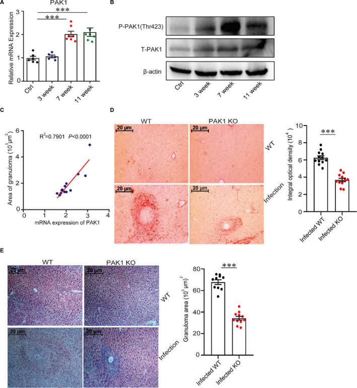
Figure 1.

PAK1 promotes hepatic pathology during S japonicum infection. (A) The PAK1 mRNA levels in the livers of WT and S japonicum‐infected mice at indicated time points post‐infection were analysed by real‐time quantitative PCR (n = 7). (B) The dynamic protein expression of phosphorylated PAK1 (Thr423) via Western blot in the livers from S japonicum‐infected mice was conducted (n = 5). (C) The correlation was shown between the granuloma size and the expression of PAK1 in the livers from S japonicum‐infected WT mice. (D, E) Paraffin‐embedded liver sections were stained with H&E or Sirius Red, the original magnification of stained liver sections was 100×, images shown were representative of experiments, scale bar, 20 μm (n = 12). Data were representative of one experiment from two replicate experiments. Data were presented as mean ± SEM. ***P < .001
