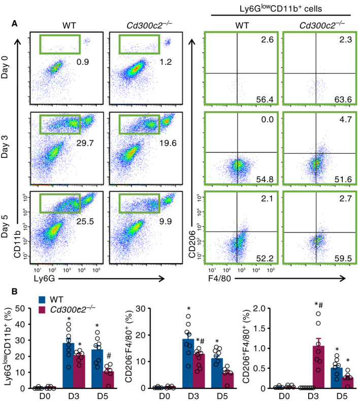
Figure 3.

MAIR‐II deficiency reduces pro‐inflammatory macrophage influx. A, Representative flow‐cytometric plots showing Ly6GlowCD11b+ macrophages, CD206‐F4/80+ pro‐inflammatory macrophages and CD206+F4/80+ anti‐inflammatory macrophages from WT and Cd300c2 −/− hearts on days 0, 3 and 5 after MI. B, Quantification of Ly6GlowCD11b+ macrophages, CD206‐F4/80+ pro‐inflammatory macrophage and CD206+F4/80+ anti‐inflammatory macrophages as a percentage of live cells from WT and Cd300c2 −/− hearts on days 0, 3 and 5 after MI. Results are presented as mean ± SEM, *P < .05 vs day 0, #P < .05 vs WT by one‐way ANOVA with Tukey's post hoc test
