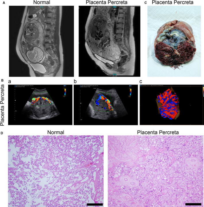
FIGURE 1.

Clinical characteristics of collected samples. (A) Magnetic resonance imaging (MRI) and (B) ultrasonic examination of normal and placenta percreta groups. The transection (a), sagittal section (b) and three‐dimensional (c) flow diagram are presented. (C) White light image of tissue samples taken during surgery. (D) Haematoxylin and eosin staining of tissue samples taken during surgery. Scale bar = 200 μm
