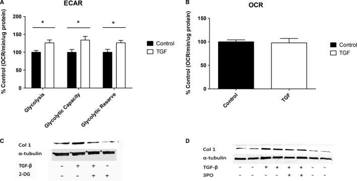
FIGURE 1.

Glycolysis is a vital component of TGF‐β1 induction of fibrotic proteins in NHDFs. A, The glycolytic parameters of NHDFs treated with/without 10 ng/mL TGF‐β1 were measured on a Seahorse XFp Analyser by performing a glycolysis stress test. B, OXPHOS was also measured in NHDFs treated with/without 10 ng/mL TGF‐β1, by measuring the oxygen consumption rate (OCR). Data shown are the percentage change compared with control untreated cells, and OCR is measured by pmol/min normalized to total protein. C, NHDFs were treated with/without 10 ng/mL TGF‐β1 and/or the glycolysis inhibitor 2‐DG (10 Mm), and the proteins indicated were measured by Western blotting. D, Likewise, Western blotting was used to observe changes to proteins of interest in NHDFs were treated with/without 10 ng/mL TGF‐β1 and/or the glycolytic flux inhibitor 3PO (8 µmol/L). *Represents P < .05. Error bars represent the mean (n = 3) ±SEM. Statistical significance was tested for using 1‐way ANOVA followed by a Bonferroni post hoc test
