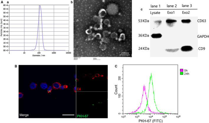
FIGURE 2.

Identification and internalization of T cell–derived exosomes. A, The size of the extracellular vesicles was tested by nanoparticle tracing assay (a, b), the morphology of exosomes was observed by transmission electron microscopy (c), and the expression of surface markers CD63 and CD9 was detected using Western blot assay (d) where 25 μg lysate of T cells (lane 1) or T cell–derived extracellular vesicles (lane 2 and 3) were loaded. B, After co‐culture for 24 h, the internalization of PKH‐67 labelled exosomes (green) by Dil labelled Jurkat cells (red) was observed by confocal microscope. Scale bar, 30 μm. C, Jurkat cells that engulfed T‐exos (PKH‐67 positive) after co‐incubation were detected by flow cytometry. Each experiment was repeated three times
