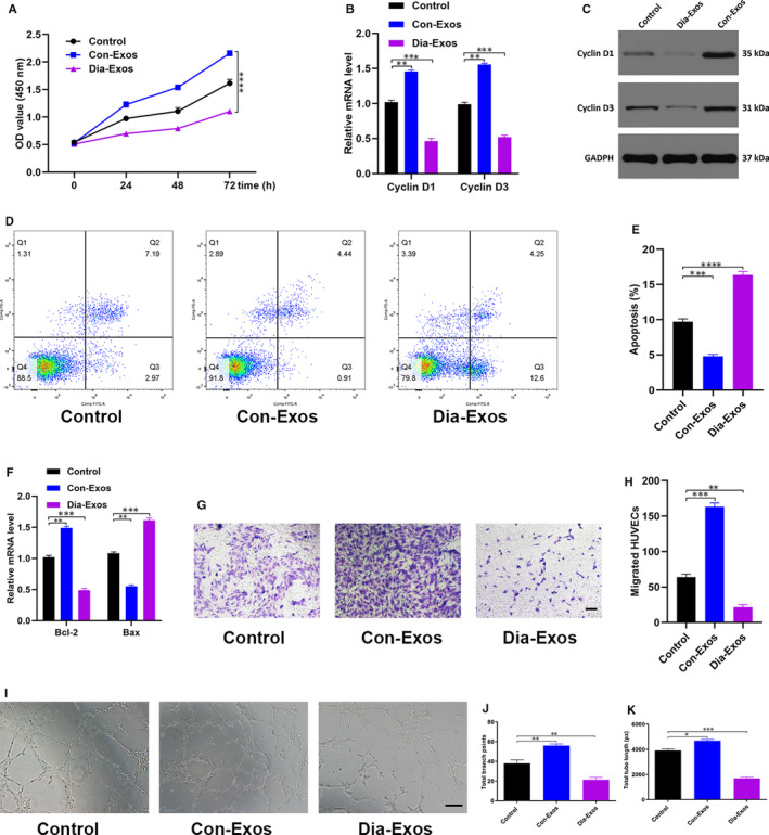
Figure 3.

Dia‐Exos inhibit HUVECs’ angiogenesis and survival in vitro. A, Effect of Dia‐Exos on HUVECs’ proliferation measured by CCK‐8 assay; (B,C) the effect of Dia‐Exos on the proliferation‐related mRNAs Cyclin D1 and Cyclin D3 levels assessed by qRT‐PCR and Western blotting; (D,E) flow cytometry was further used to quantify cell apoptosis, showing a greater percentage of HUVECs was in G1 phase and apoptotic following Dia‐Exos treatment; (F) the effect of Dia‐Exos on the apoptosis‐related mRNAs Bcl‐2 and Bax levels assessed by qRT‐PCR analysis; (G,H) the effect of Dia‐Exos on HUVEC migration was assessed by transwell migration assay, scale bar, 100 µm; (I‐K) effects of Dia‐Exos on the angiogenesis ability of HUVECs was measured by tube formation assay, scale bar, 200 µm. Data are the means ± SD three independent experiments. *P < .05, **P < .01, ***P < .001
