
Figure 5.
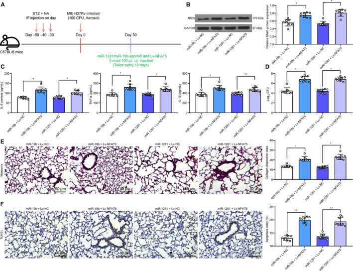
Lv‐NFAT5 partially blocks the effects of miR‐19b/miR‐1281 agomiR. A, 1 mo after diabetes induction, mice were exposed to ~100 CFU of aerosolized Mtb; another 30 d later, each mouse was further given miR‐19b/1281 agomiR, or the LV overexpressing NFAT5 (5 nmol/100 μL) twice every 10 d through i.p for a total of 30 d; B, protein level of NFAT5 in mouse lung tissues determined by Western blot analysis; C, secretion of pro‐inflammatory factors in mouse lung tissue homogenate determined by ELISA kits; D, Mtb content in lung tissues; E, lung fibrosis in mouse lung tissues determined by Masson's trichrome staining; F, apoptosis in lung tissues determined by TUNEL. Each spot in the images indicates one sample. N = 8 in each group Three independent experiments were performed. Data were expressed as mean ± SD compared by one‐way ANOVA and Tukey's multiple comparison test. *P < .05, **P < .01