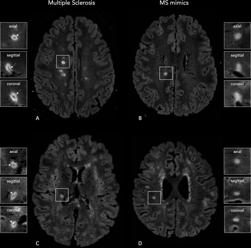
Figure 1 -. 3D FLAIR* images obtained on Siemens (Lausanne University Hospital; A,B) and Philips (Erasmus University Hospital; C,D) MRI scanners showing central vein sign positive (CVS+) and negative (CVS-) lesions.
A central vein running through the lesion (arrows) is visible in the majority of white matter lesions in a 28-year-old man (A) and 27-year-old woman (C) with relapsing-remitting MS. Images from 25-year-old (B) and 46-year-old (D) women with Sjögren syndrome show how the central vein sign is not typical of white matter lesions in MS-mimicking diseases.

