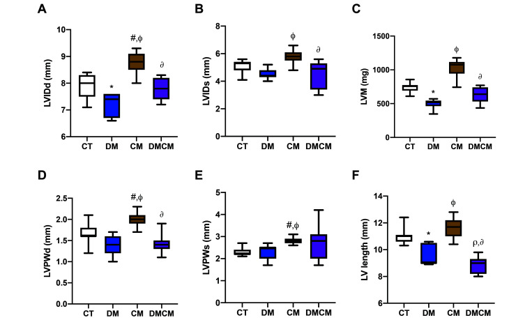
Figure 2.
Ventricular dimension and mass indices with (A) LVIDd left ventricular internal diameter at end diastole, (B) LVIDs left ventricular internal diameter at systole, (C) LVM left ventricular mass, (D) LVPWd left ventricular posterior wall thickness at end diastole, (E) LVPWs left ventricular posterior wall at systole, and (F) LV length left ventricular length (measured from closed mitral valve (early systole) to apex in apical 4-chamber view). CT control group, DM diabetic group, cardiomyopathy cardiomyopathic group, DMCM diabetic, and cardiomyopathic group. *P < 0.05 CT compared with DM,#P < 0.05 CT compared with CM,rP < 0.05 CT compared with DMCM,fP < 0.05 DM compared with CM,∆P < 0.05 DM compared with DMCM,∂P < 0.05 CM compared with DMCM

