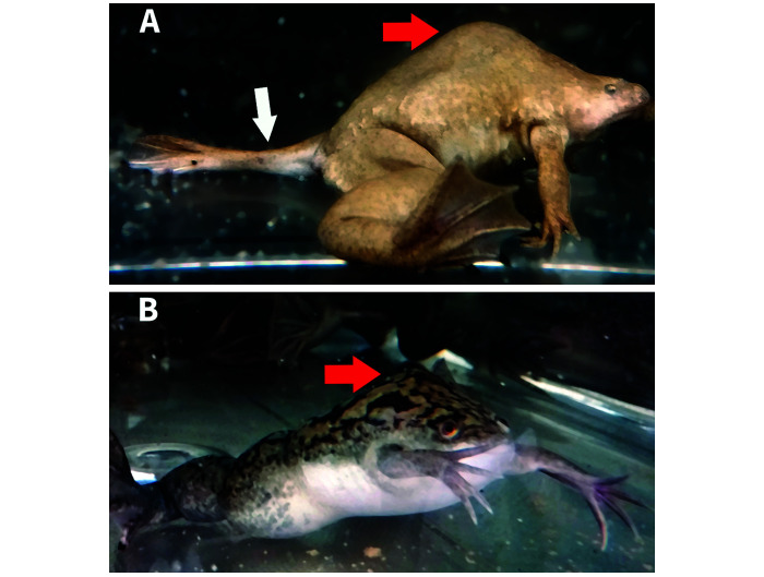
Figure 2.
Two frogs displaying dorsal humps (red arrows) indicative of vertebral malformation, which represent this cohort's affected condition. Lateral view (enlarged 2×) of affected X. tropicalis (A) shows kyphosis and the abnormal positioning of the left rear leg due to coxofemoral subluxation (white arrow). Lateral view of an affected X. laevis (B). No other skeletal abnormalities grossly visible.

