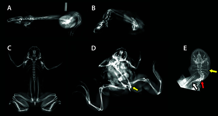
Figure 4.
Right lateral radiographs of a control X. laevis (SVL approximately 110 mm) (A) and an affected X. laevis (SVL approximately 50 mm) (B) with severe kyphosis. Ventrodorsal view of a control X. laevis (C). Ventrodorsal views of an affected X. laevis showing scoliosis and coxofemoral luxation (yellow arrow) (D), and another affected X. laevis showing scoliosis focused in the presacral vertebrae (red arrow), with misshapen transverse processes (yellow arrow) and a curved urostyle (white arrow). The normal appendages are not shown on the X. laevis in E to enhance radiographic details that were otherwise difficult to discern.

