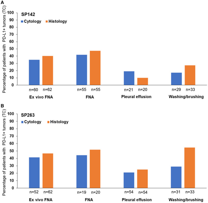
FIGURE 3.

Histograms representing the percentages of patients with TC PD‐L1 expression in histology and cytology samples according to the cytological sample type with (A) the SP142 antibody and (B) the SP263 antibody. FNA also included transbronchial needle aspiration. There were differences in the numbers of evaluable histology and cytology samples between the SP142 and SP263 results. Positive PD‐L1 expression (PD‐L1+) was defined as ≥1% of TC. FNA indicates fine‐needle aspiration; PD‐L1, programmed death ligand 1; TC, tumor cell.
