
FIGURE 5.
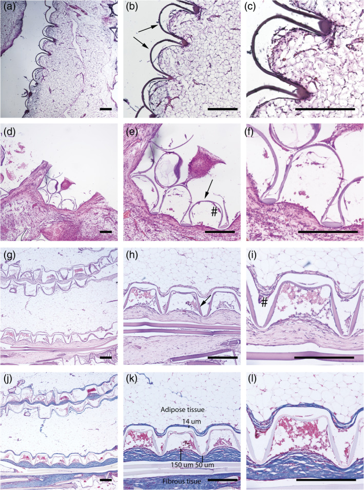
Hematoxylin eosin staining of intraperitoneally implanted 4000PEOT30PBT70 rats after week 1 (a)–(c), week 4 (d)–(f) and week 12 (g)–(i), and trichrome staining of week 12 samples (j)–(l) with blue stain for collagen, red/pink for cellular cytoplasm and black for nuclei. The presence of foreign body giant cells (#) is limited. Scale bar in all light micrographs indicates 300 μm