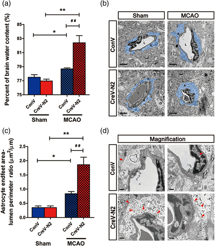
FIGURE 1.

NDRG2 deficiency exacerbates brain edema and astrocyte swelling after stroke. (a) Quantification of brain water content in cortices isolated from Ndrg2 GFAP cKO mice (CreV‐N2) and control mice (ConV) in sham groups and MCAO groups. Data are shown as the mean ± SD (n = 5–6). *p < .05, **p < .01 versus sham mice, ## p < .01 versus control mice. (b) Representative transmission electron micrographs showing the swollen perivascular astrocytic end‐feet area (blue area). A: astrocyte end‐feet; B: basal lamina; E: endothelial cell; L: lumen. Scale bar = 2 μm. (c) Astrocyte end‐feet area‐to‐capillary lumen perimeter ratio. Data are shown as the mean ± SD (n = 5–6). *p < .05, **p < .01 versus sham mice, ## p < .01 versus control mice. (d) High‐magnification images of ultrastructural changes in MCAO group mice. Red arrowheads indicate disruption of the plasma membrane, basal membrane, gap junctions, organelles, accumulation of glycogen, and swollen endothelial cytoplasm. Scale bar = 0.5 μm [Color figure can be viewed at wileyonlinelibrary.com]
