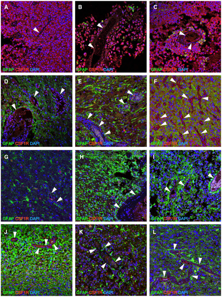
Figure 5.

Double immunohistochemistry for CSF‐1R and GFAP. When CSF‐1R expression was compared to that of the vessels (arrow head), which was used as an internal positive control of CSF‐1R, strong and diffuse expression was detectable in all E‐GBM tumor cells [CSF‐1R (Cases 1–3; A–C: red) and DAPI (A–C: blue)]. By contrast, although CSF‐1R expression was observed in PXA [CSF‐1R (Cases 4 and 5; D, E: red) and DAPI (D, E: blue)] and A‐PXA [CSF‐1R (Case 6; F: red) and DAPI (F: blue)], its expression was not stronger than that of the vessels, and it was detectable only regionally. Similarly, in conventional GBM [CSF‐1R (Cases 7–10, 12; G–J, L: red) and DAPI (G–J, L: blue)] and AA [CSF‐1R (Case 11; K: red) and DAPI (K: blue)], CSF‐1R expression was not stronger than that in the vessels. No GFAP+ cells [GFAP (Case 1; A: green)] or only a few GFAP+ cells [GFAP (Cases 2 and 3; B, C: green)] were detectable in E‐GBM, whereas many GFAP+ cells were observed in PXA [Cases 4 and 5; GFAP (D, E: green)], A‐PXA [Case 6; GFAP (F: green)], conventional IDH‐WT GBM [Cases 7–10; GFAP (G–J: green)], IDH‐MT AA [GFAP (Case 11; K: green)] and IDH‐MT GBM [GFAP (Case 12; L: green)] .Abbreviations: AA, anaplastic astrocytoma; A‐PXA, atypical pleomorphic xanthoastrocytoma; CSF‐1R, colony‐stimulating factor 1 receptor; DAPI, 4′,6‐diamidino‐2‐phenylindole; E‐GBM, epithelioid glioblastoma; GBM, glioblastoma; GFAP, glial fibrillary acidic protein; MT, mutant; PXA, pleomorphic xanthoastrocytoma; WT, wild‐type.
