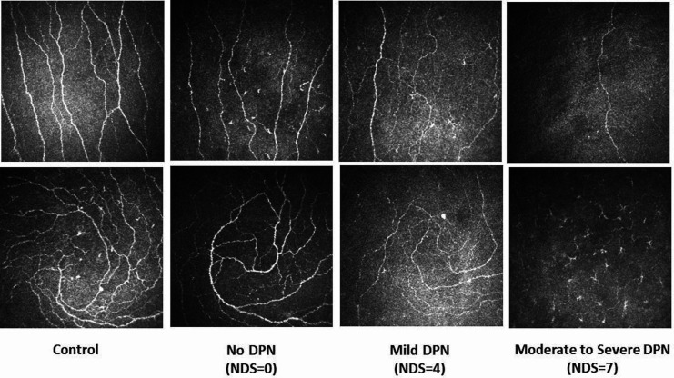
Figure 1.
Corneal confocal microscopy images of the central cornea (top row) and the inferior whorl (bottom row) in a healthy control (first column) and in patients with no (second column), mild (third column) and moderate to severe (fourth column) neuropathy. DPN, diabetic peripheral neuropathy; NDS, Neuropathy Disability Score.

